Columbia neuroscientists have discovered why mitochondria, tiny power generators that keep our cells healthy, are often strangely shaped inside the brain. Mitochondria, which exist by the thousands in each of our body’s 37 trillion cells, usually look like long interconnected tubes. But inside brain cells called neurons, they adopt two completely different shapes depending on their location within the cell: that same elongated, tubular shape and a substantially smaller, almost spherical shape, that more closely resembles golf balls. In today’s study, researchers have identified the mechanism responsible for these differences in mitochondrial shape — uncovering key insight into the relationship between mitochondrial shape and their function.
This research, published online today in Nature Communications, suggests that these unusually small, squat mitochondria help neurons grow and make proper connections in the developing brain. The work could open up new lines of inquiry into may be at play when these processes go awry in brain disease.
Not delving into the inner workings of the brain at this level of detail would be akin to trying to understand how a car works just by watching it move along a highway. You have to open up the car’s hood and take a close look at all its parts.
“In most cells in our body, mitochondria take on the standard tubular shape, but inside neurons, mitochondria can adopt that same shape or they can be tiny, almost spherical; the reasons for this difference have remained largely unexplored,” said Franck Polleux, PhD, a principal investigator at Columbia’s Mortimer B. Zuckerman Mind Brain Behavior Institute and the paper’s senior author. “Today, we’ve uncovered an unexpected mechanism that helps maintain the unusually small size of these mitochondria, shedding new light on how maintenance of their size is critical for normal brain-cell growth.”
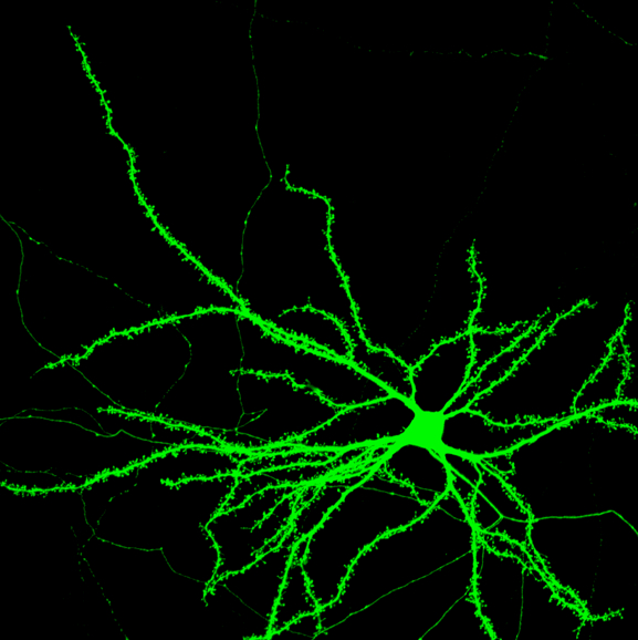
The unique shape of these mitochondria is ultimately tied to the unique shape of neurons themselves. Unlike other cells in the body that are comprised only of a simple cell body, neurons also have two sets of extensions, branching outward in opposite directions, called dendrites and axons. These extensions are essential. They span out from the neuron like tendrils, linking up to other cells to pass information to each other in the form electrical pulses — forming an information superhighway of microscopic proportions.
To pass these pulses, an axon from one neuron will connect to the dendrite of another neuron. That contact point, called a synapse, acts like an intercellular ‘handshake’ to ensure the right electrical pulses — and therefore the right information — are shared between cells.
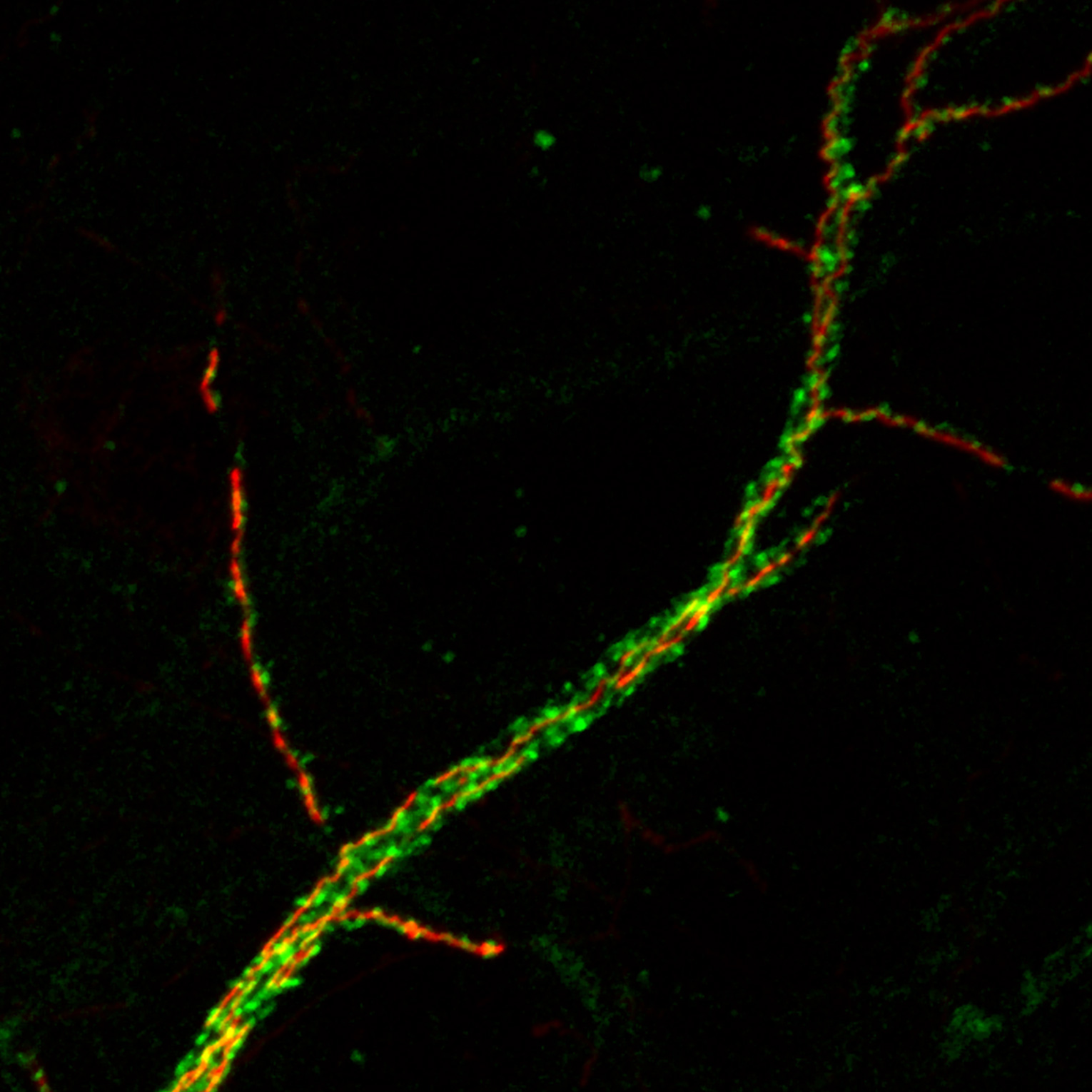
To bolster cellular communication, a single axon can form thousands of individual branches, each of which is about one micron in diameter — 50 times smaller than the width of a human hair. In each axon and its branches exist thousands of small mitochondria, which are often localized at synapses.
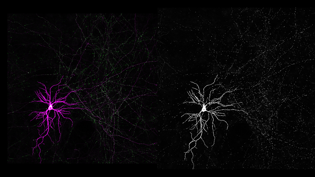
Left: a neuron with all its synapses shown in green. Right: that same neuron with its mitochondria shown in white (Credit: Tommy Lewis/Polleux lab/Columbia's Zuckerman Institute).
“These axonal mitochondria are unlike any mitochondria anywhere else in the body — they are even different than those found in other parts of the neuron,” said Dr. Polleux, who is also a professor of neuroscience at Columbia University Irving Medical Center. “This begged the question: Does this small size serve a function?”
Mitochondria are remarkably dynamic. In order to keep their size uniform, they constantly undergo fusion (in which several mitochondria will combine into one) and fission (in which they split apart). In a series of experiments on neurons taken from the brains of mice, the researchers, including co-first author and former Polleux lab member Tommy Lewis, PhD, pinpointed a gene called MFF. When switched on, MFF appears to keep axonal mitochondria decidedly small.
“The gene MFF promotes mitochondrial fission,” said Dr. Polleux. “so, when we shut off MFF, we tilted the balance, increasing fusion events. This caused the normally small axonal mitochondria to increase in length by five-to-ten-fold.”
Surprisingly, this increase in size did not reduce mitochondria’s ability to move up and down the axon. Nor did it change their capacity to act as energy powerhouses, which was wholly unexpected, given that this is thought to be their main role in most cells.
There was one significant difference in these new, longer axonal mitochondria, uncovered by co-first author and former Polleux lab member Seok-Kyu Kwon, PhD: They took in a significantly greater amount of calcium from their surroundings. Calcium is critical for brain activity, including for transmitting between synapses. Mitochondria’s calcium uptake normally allows electrical signals to pass between cells.
But longer mitochondria meant a greater capacity to take up calcium at synapses. This, the researchers observed, disrupted the normal pattern of electrical signals that passed between cells, impairing the neurons’ ability to communicate with their neighbors. This impairment also stunted the axon during development, ultimately causing them to decrease their branching.
“The normally small size of axonal mitochondria seems to keep calcium buffering at just the right level to drive healthy axonal growth and foster cellular connections,” said Dr. Polleux.
Interestingly, these findings also suggest that energy production — the primary job of mitochondria elsewhere in the body — may not be the main duty of axonal mitochondria.
“There may be a far more specialized role for them,” said Dr. Polleux. “And that is something that we are actively deciphering at this very moment.”
For Dr. Polleux, knowing the steps that drive neuronal growth is a critical step toward untangling neurodegenerative disease. Many diseases, including Alzheimer’s, affect mitochondrial structure and function. He hopes the work in his lab can help shed light on why this is the case, and also what steps can be taken to mitigate it.
“Not delving into the inner workings of the brain at this level of detail would be akin to trying to understand how a car works just by watching it move along a highway,” he added. “You have to open up the car’s hood and take a close look at all its parts.”
###
This paper is titled “MFF-dependent mitochondrial fission regulates presynaptic release and axon branching by limiting axonal mitochondria size.” Additional contributors include Annie Lee, PhD (Columbia University) and Reuben Shaw, PhD (Salk Institute).
This research was supported by the National Institutes of Health (R01NS089456, K99NS091526), the Human Frontiers Science Program and the Fondation Roger de Spoelberch.
The authors report no financial or other conflicts of interest.