Your body is comprised of many trillions of cells. But it began with just one. The steps required to complete that journey — from one cell to trillions — have long been a subject of intense study by scientists across disciplines.
One group of proteins that has an especially important role in this process are called Notch proteins. These proteins are everywhere; in the majority of cells of most animals on the planet. Their pervasiveness speaks to their pivotal role in biological life: These proteins guide cells, young and old, to make decisions about their ‘cellular identity’ — who they are and what they will become.
Yet despite their ubiquity across cells, and species, as well as the myriad roles they are known to play in growth and development, there has been fierce scientific debate about how Notch proteins function, and especially the steps required to ‘switch on’ or activate them in the first place.
Research has shown that this process is far more complicated than flipping a switch. Instead, it involves a coordinated sequence of steps — like a microscopic Rube-Goldberg device. The challenge for scientists has been tracing the key events in this sequence.
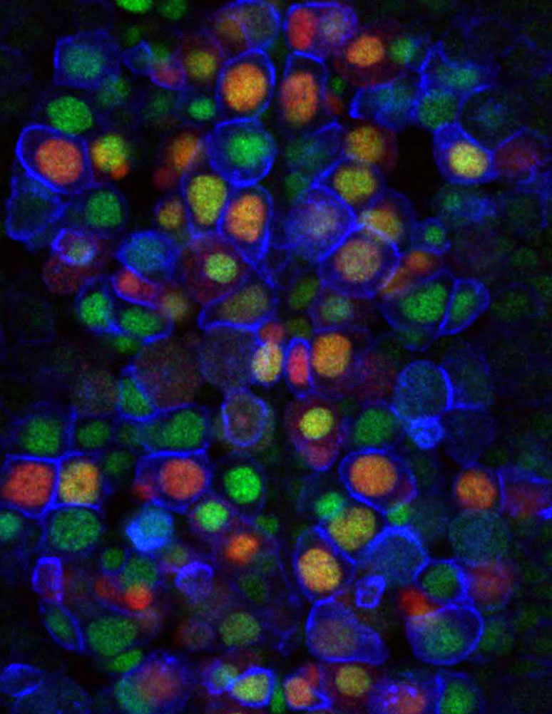
But in new research published this week in Cell, scientists at Columbia’s Mortimer B. Zuckerman Mind Brain Behavior Institute have devised powerful new cell-to-cell signaling technologies that do precisely that, called Mosaic Analysis by Promoter Swap, or MAPS.
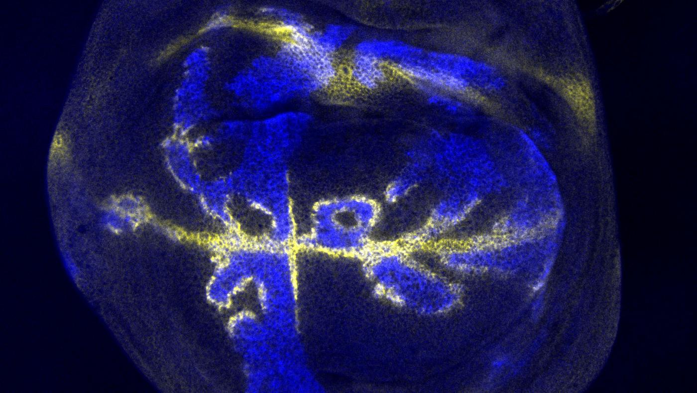
Because we could do all this in a developing fruit fly — as opposed to isolated cells in a petri dish — we got a front-row seat to view one of biology’s most ancient and important systems.
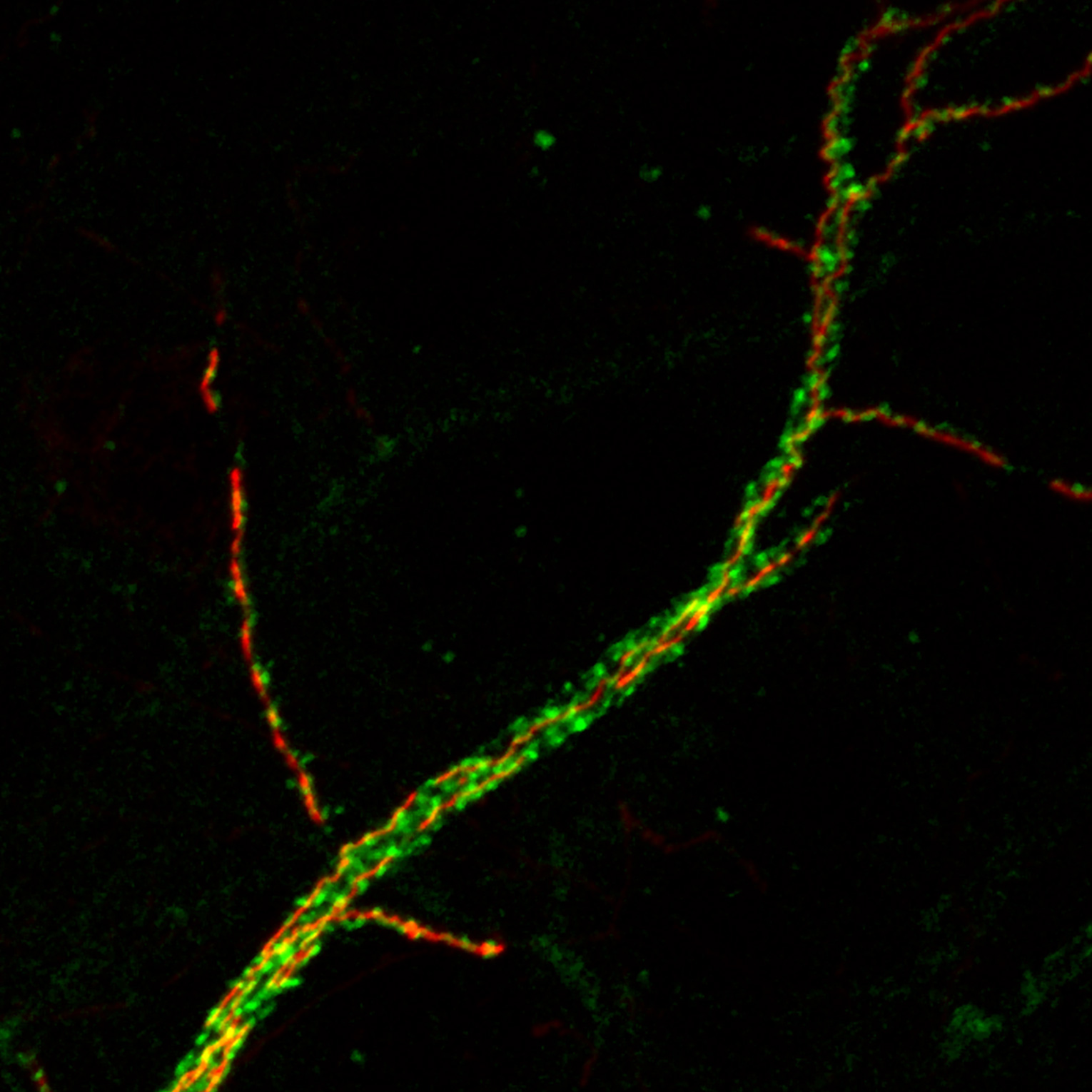
Developed by Zuckerman Institute Principal Investigator Gary Struhl, PhD, and Paul Langridge, PhD, an associate research scientist in his lab, MAPS lets scientists interrogate what is known as S2 cleavage, one of the earliest and key steps along the complex Notch activation sequence. In S2 cleavage, a portion of Notch protein is literally cut off, or ‘cleaved,’ from the surface of a developing cell. The remaining segment of protein is then free to go the cell nucleus and activate a group of genes that are known to control the cell’s fate. But what triggers this cleavage has remained unsolved.
This is where MAPS comes in.
“MAPS gave us the power to monitor, and even control, the interaction between Notch and other proteins involved in the S2 cleavage,” said Dr. Langridge, who served as the paper’s first author. “And because we could do all this in a developing fruit fly — as opposed to isolated cells in a petri dish — we got a front-row seat to view one of biology’s most ancient and important systems.”
Strikingly, MAPS allowed the researchers to decipher how S2 cleavage works: Other proteins, sitting on the surfaces of neighboring cells, exert a physical force on the Notch protein, which induces S2 cleavage. Surprisingly, MAPS further revealed that this force is generated when neighboring cells internalize the proteins on their surface, a process known as endocytosis. Endocytosis of these proteins, which are bound to Notch proteins that are located on the abutting cell surface, exerts mechanical tension on the Notch protein. This ‘pulling’ effect causes this outermost section of Notch to stretch open, exposing an otherwise hidden site that can then be cleaved.
These findings, while lending critical insight into S2 cleavage, could also help to boost understanding of Notch’s role in development more broadly. For example, dysfunctions in Notch have been implicated in a wide range of diseases, from developmental disorders to cancer. MAPS could one day be applied to further scientific understanding of the mechanisms behind these dysfunctions.
“Furthermore, recent advances in synthetic biology have led to the development of Notch-like artificial proteins that could serve as a therapeutic intervention of disease at the cellular level,” added Dr. Langridge. “Our work provides essential parameters for adapting and optimizing these so-called synthetic notch, or ‘Syn-Notch,’ cellular circuits for potential use in therapeutics.”
###
This paper is titled: “Epsin-dependent ligand endocytosis activates notch by force.”
This research was supported by the National Institutes of Health (R01 GM109183, P400D018537) and the Howard Hughes Medical Institute.
The authors report no financial or other conflicts of interest.