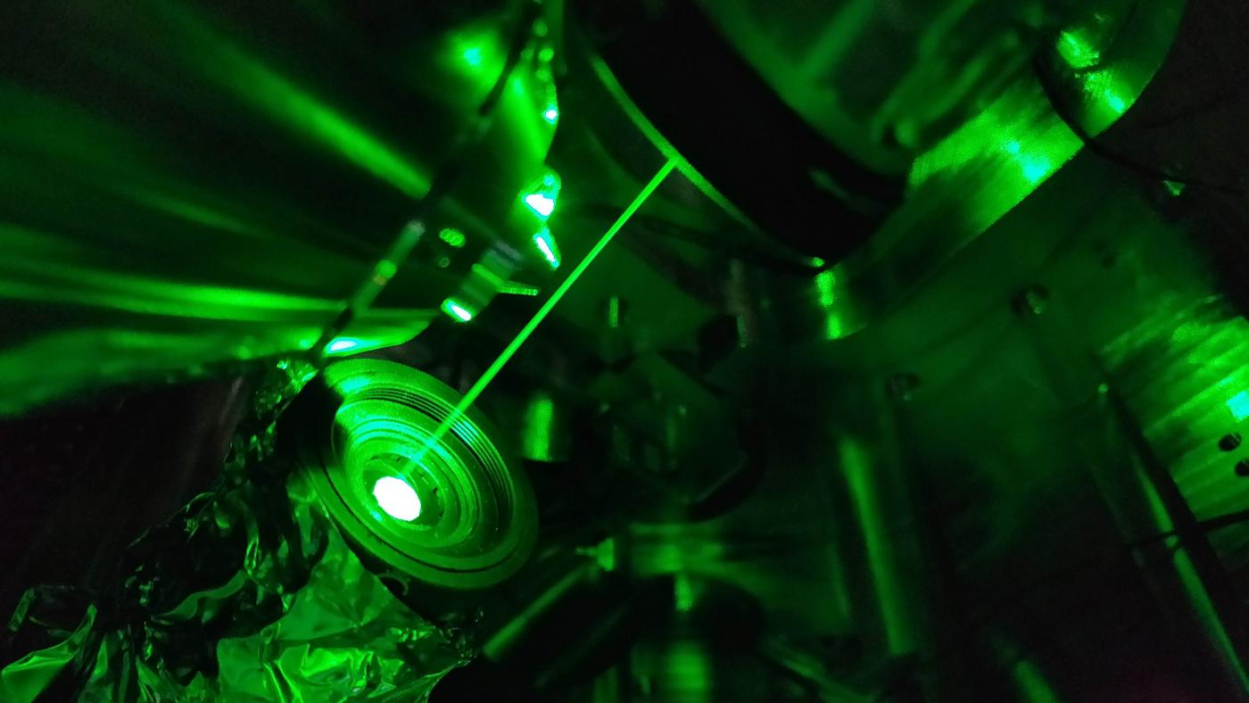
NEW YORK, NY — The laser you see in this photo may one day enhance images taken by the most powerful microscopes in biology. This advance, detailed in eLife, from scientists at Columbia's Zuckerman Institute with the Maxson lab at Cornell University, could revolutionize research into the molecules that allow the brain to function properly and underlie diseases.
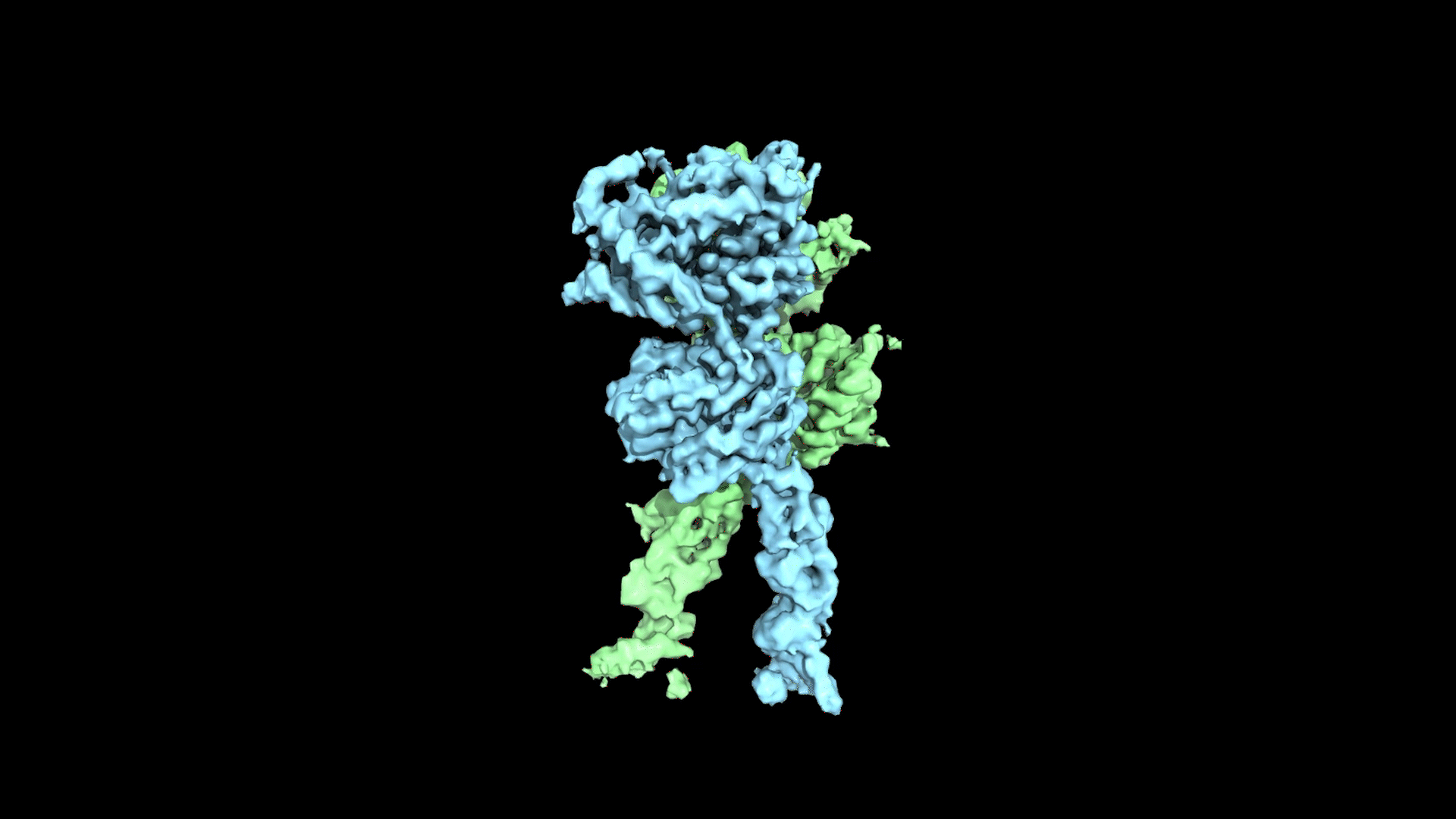
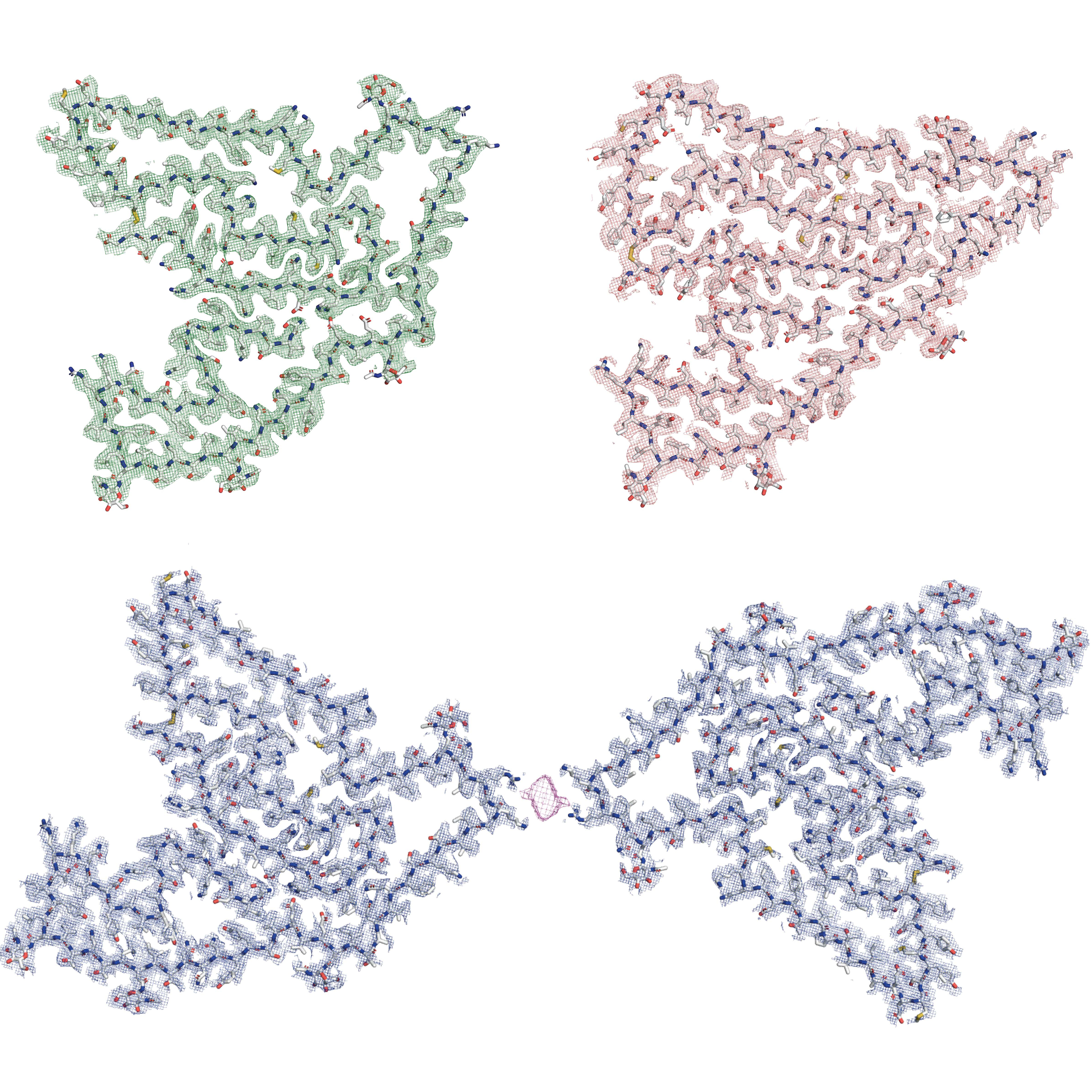
Cryo-electron tomography (cryo-ET) fires beams of electrons at frozen specimens. This enables researchers to create 3D images of molecules in the samples. Electrons accelerated to high speeds have a smaller wavelength than the visible light used in conventional microscopes, so cryo-ET can generate far more detailed images, with near-atomic-level resolution.
"Electron microscopy can help us visualize what’s going on in the synapse, a gap only 20 billionths of a meter wide where neurons connect and communicate," says Anthony Fitzpatrick, PhD, a principal investigator at Columbia’s Zuckerman Institute, the study's senior author and an assistant professor biochemistry and molecular biophysics at Columbia’s Vagelos College of Physicians and Surgeons.
Having a detailed picture of where and how these molecules interact could help us find new ways to deal with them
Using cryo-ET, the Fitzpatrick lab is exploring how molecules linked to Alzheimer’s, Parkinson’s and other devastating diseases travel inside cells. "Having a detailed picture of where and how these molecules interact could help us find new ways to deal with them," says Daniel Du, PhD, the study’s lead author and a postdoctoral scientist in the lab.
As powerful as cryo-ET is, one limitation it faces is how weakly biological molecules interact with electrons. This leads to low-contrast, washed-out images. In the new study partially funded by the Chan-Zuckerberg Initiative, the team worked with Tanya Tabachnik and the institute’s Advanced Instrumentation team to connect a laser to a proof-of-concept microscope. Their initial research confirmed they could use their laser to target electrons traveling at samples. Their ultimate goal is for the laser to target electrons that pass through samples without interacting. This will change the properties of those electrons, helping boost contrast and greatly improve the pictures.
"To make this work, you have about a trillionth of a second to hit, with a laser, an electron that's traveling at almost the speed of light," Dr. Du says. "How cool is that?"