Columbia scientists have uncovered a key feature of the brain’s GPS that helps a mouse find what it is seeking. The study enabled scientists to define the precise duties of cells in a particular region of the hippocampus, the brain’s learning and memory center. The research also advances a long-standing quest in the field of neuroscience: tracing the pathway that information takes while traveling through the brain.
The authors announced these findings today in the journal Neuron.
“In this study, our goal was to simulate what our brains do as we walk aimlessly down the street, versus how our brains behave when looking for a specific address,” said Attila Losonczy, MD, PhD, a principal investigator at Columbia’s Mortimer B. Zuckerman Mind Brain Behavior Institute, associate professor of neuroscience at Columbia University Medical Center (CUMC) and the paper’s senior author. “By using the powerful two-photon microscope, we were able to observe the activity of individual cells in the mouse hippocampus, and then link that activity to a specific behavior — in this case, navigation — a technological feat that would have been impossible just a few years ago.”
It’s astounding that the ability to navigate to a desired location, an enormously complex feat, can be represented so precisely in the structure of the hippocampus.
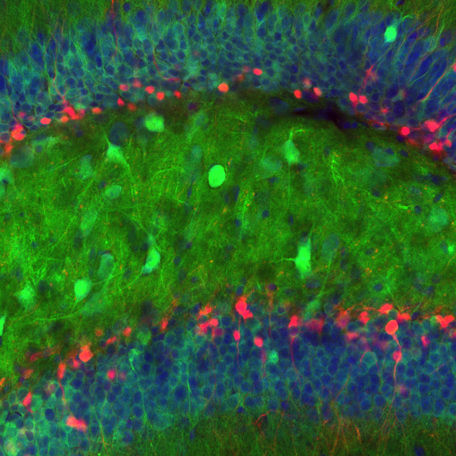
The hippocampus can be divided into distinct areas that form an interconnected circuit through which memory-related information is processed. For this study, Dr. Losonczy and his team focused on the hippocampus’ main output node, area CA1, which was discovered by scientists to encode one’s location — work that was awarded the 2014 Nobel Prize.
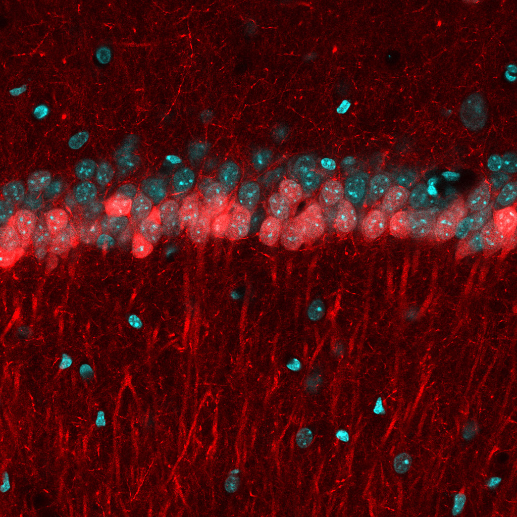
Above: Microscopic image of cells in the CA1 region of the hippocampus, divided into two sublayers. Cells in the superficial layer are stained red, while cells in the deep sublayer are stained in cyan (greenish-blue)(Credit: Nathan Danielson/Columbia's Zuckerman Institute).
“We’ve known that CA1 can be divided into two distinct sublayers of cells: the deep and superficial sublayers,” said Nathan Danielson, a doctoral candidate in neuroscience at CUMC and the paper’s first author. “Scientists have wondered whether this division was an indication that these two sublayers actually served different purposes in learning and memory. But no one had tested it, so we decided to look.”
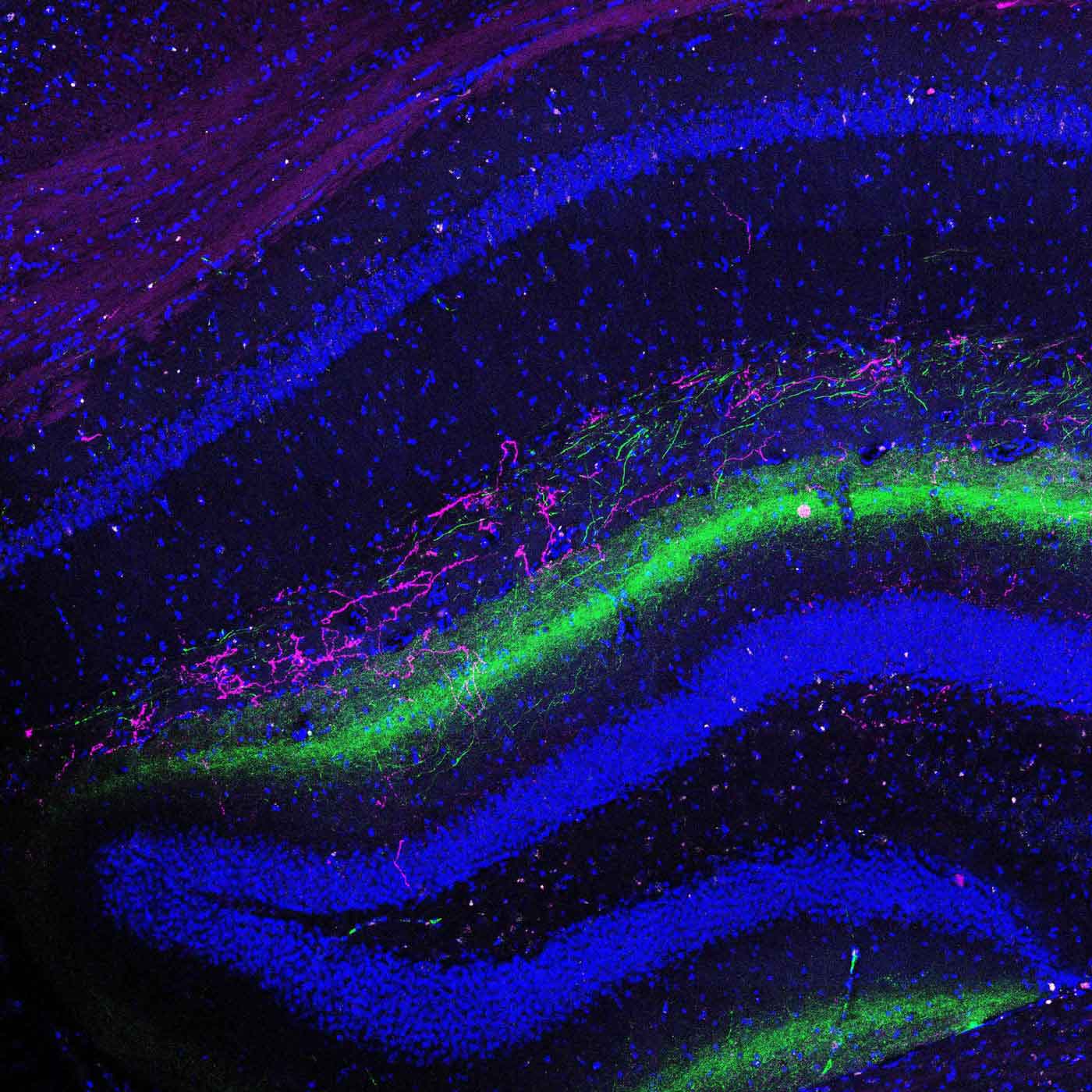
To study these cells, the researchers placed mice on treadmills that had distinct colors, textures and smells while a two-photon microscope monitored the cellular activity in the CA1. The mice then performed two tasks.
In the first, mice ran on a treadmill while experiencing different sights and sounds, some familiar and others new. In the second, mice were given the task of finding a water reward placed at a specific, unmarked location along the treadmill. The team repeated these experiments over the course of several sessions and monitored how each of the sublayers responded to the different types of learning.
When the mice performed the first task, cells in the superficial sublayer of CA1 appeared to create an internal map that remained largely unchanged from session to session. By contrast, cells in the deep sublayer formed an internal map that was far more dynamic — in effect redrawing a different version of the map during each session.
During the second task, however, when the mice needed to learn the location of the hidden reward, the maps in the deep sublayer were significantly more stable, and less dynamic, than in the first task. The scientists also found that deep-sublayer activity was closely linked to the animal’s ability to find the reward. This distinction between the sublayers, the authors argue, could signify two different processes important for navigation.
“If you’re walking down the street looking for something specific — say, your favorite restaurant — your brain first needs a map of the neighborhood in general,” said Danielson. But to find that particular restaurant, he continued, the brain also assigns importance, or salience, to that specific location.
“In a sense, it’s the brain’s way of marking a location on a map with a giant X,” Danielson said. “So as you look for that restaurant, you need both the map and the X. Our findings suggest that, in the brain, these distinct types of information could be conveyed by the CA1’s distinct sublayers.”
“And if one month later you wanted to visit somewhere new, the deep sublayer would update the map, effectively marking the spot of the new location, while the underlying map of the neighborhood, created by the superficial sublayer, would remain relatively unchanged,” added Dr. Losonczy.
For Dr. Losonczy, this study speaks to the ingenious way that the brain’s underlying architecture allows it to accomplish a specific type of navigation.
“It’s astounding that the ability to navigate to a desired location, an enormously complex feat, can be represented so precisely in the structure of the hippocampus,” he said. “And it’s even more astounding that we can now witness it happening in real time.”
###
This paper is titled: “Sublayer-specific coding dynamics during spatial navigation and learning in hippocampal area CA1.” Additional contributors include Jeffrey Zaremba, Patrick Kaifosh, PhD, John Bowler and Max Ladow.
This research was supported by the National Institute of Neurological Disorders and Stroke (F30NS090819), the National Institute of Mental Health (91F31MH105169, 1R01MH100631, 1U01NS090583, 1R01NS094668), the Howard Hughes Medical Institute, the Searle Scholars Program, the Human Frontier Science Program and the McKnight Foundation.
The authors report no financial or other conflicts of interest.