Columbia scientists have found that spikes in the activity of neurons in young mice do not spur corresponding boosts in blood flow — a discovery that stands in stark contrast to the adult mouse brain. This new study raises questions about how the growing human brain meets its energy needs, as well as how best to track brain development with functional magnetic resonance imaging, or fMRI, which relies on blood-flow changes to map neuronal activity in the brain. The research could also provide critical new insights for improving care for infants.
The findings were published today in the Journal of Neuroscience.
“In the adult brain, neuronal activity triggers a localized boost in blood flow. This relationship between neuronal activity and blood flow has long been assumed to be present from birth, but our findings in mice suggest the opposite: instead, it develops over time,” said Elizabeth Hillman, PhD, a principal investigator at Columbia’s Mortimer B. Zuckerman Mind Brain Behavior Institute, associate professor of biomedical engineering and radiology at Columbia’s Fu Foundation School of Engineering and Applied Science and the paper’s senior author. “Our study further suggests that this process is an essential part of building a healthy brain and could represent an unexplored factor in brain disorders that emerge in early childhood.”
Today’s study was motivated by previous fMRI studies in humans that reported vastly different responses in the brains of babies compared to the brains of adults.
“No one knew how to interpret blood-flow responses in the developing brain,” said Mariel Kozberg MD, PhD, a recent Columbia neurobiology graduate in Dr. Hillman’s lab and the paper’s first author. “In this study, we needed to find out what was different between adult and newborn brains. Were the differences in neural activity itself, or did they lie in the relationship between this activity and local blood flow changes?”
To answer this question, Drs. Hillman and Kozberg developed a new imaging technique that simultaneously recorded neuronal activity and blood flow in the brains of mice of different ages (from newborn up to adult), tracking how the brain responded when they stimulated each animal’s hind paw.
“When we started to get data we were amazed by what we could see,” said Dr. Kozberg. First, the team’s innovative imaging methods revealed that, for the youngest mice, stimulating the hind paw caused a strong neuronal response, but this response was localized to one region. Then, as the animals got older, the neuronal response began to spread. By 10 days of age, stimulating the right paw first sparked activity on the left side of the brain before traveling to the right side, corresponding to the development of connections between the two hemispheres.
“We realized we were actually watching cells form connections with each other throughout the brain: the development of neural networks,” added Dr. Hillman, who is also a member of the Kavli Institute for Brain Science at Columbia.
The researchers’ second finding was even more startling. In the youngest mice, neuronal activity did not trigger an increase in blood flow, as occurs in the adult mouse brain. But as the animals matured, and their neural networks became more established, the brain’s blood-flow response gradually got stronger over time until the animal reached adulthood.
“It was like the brain was gradually learning to feed itself,” said Dr. Hillman, who notes that this finding makes a lot of sense. “It is hardly surprising that blood vessels — and the machinery linking them to brain activity — would mature in step with the development of neural activity itself.”
However, these results raised a worrying question. The job of blood vessels is to deliver oxygen-rich blood to the brain. So, can the newborn brain truly function and grow without subsequent increases in blood flow? To find out, Drs. Kozberg and Hillman used another optical imaging technique, called flavoprotein imaging, which measures how the newborn brain used oxygen.
Neural activity in the newborn brain might actually be guiding blood vessels to grow in the right places.
“In the youngest animals, we confirmed that neurons were indeed consuming oxygen, but without a rush of fresh blood, they seemed to run out of fuel,” said Dr. Kozberg. “We further found that the neural activity actually caused localized drops in oxygen levels, known as hypoxias.”
Drs. Hillman and Kozberg propose several explanations for this surprising result. “Newborns make an incredible transition from being inside the womb to breathing air in the delivery room,” noted Dr. Hillman. “To survive those first few hours, the newborn brain must be well prepared to withstand enormous fluctuations in the availability of oxygen.”
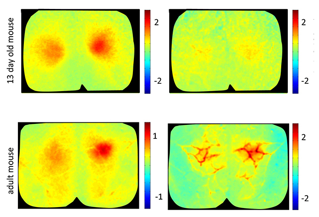
Brain activity (left) associated with more blood flow (right) in adult mice (bottom), but not in young mice (top) (Credit: Lab of Elizabeth Hillman, PhD, principal investigator at Columbia's Zuckerman Institute).
Because the hypoxias seen in young mice appear to be part of the normal development process, the authors propose that it may in fact serve an important purpose.
“We know that a lack of oxygen can trigger the growth of blood vessels,” said Dr. Kozberg. “So in this case, neural activity in the newborn brain might actually be guiding blood vessels to grow in the right places.”
Moving forward, the team is preparing to compare their results in mice to the human brain. Dr. Hillman is working with researchers in Columbia’s Department of Psychiatry to analyze hundreds of fMRI scans previously collected from newborns, as well as from children of different ages.
“If we can find the same signatures of neurovascular development in human infants, we could turn fMRI into an even more powerful tool. For example, using it to better understand, detect and track the origins of developmental disorders in the newborn brain,” she said.
Dr. Hillman’s team is also eager to continue studying oxygen metabolism in newborns. Preterm infants exposed to high oxygen levels can suffer from retinopathy, a condition in which blood vessels in the eyes grow incorrectly. She hypothesizes that excessive oxygen could lead to the same disruptions to blood-vessel growth in the brain itself.
Added Dr. Hillman, “If we can learn more about the unique metabolic state of the developing brain, we might be able to improve treatment strategies for premature infants, while also gaining a deeper understanding of normal and abnormal brain development overall.”
###
This paper is titled: “Rapid postnatal expansion of neural networks occurs in an environment of altered neurovascular and neurometabolic coupling.” Additional co-authors include Ying Ma, Mohammed A. Shaik and Sharon H. Kim.
This research was supported by The National Institutes of Health (1R01NS063226, 1R01NS076628, R21NS053584, T32 GM07367 MSTP, F31 NS084538), the National Science Foundation (CAREER 0954796), the Human Frontier Science Program and the Kavli Foundation.
The authors report no financial or other conflicts of interest.