Herbert and Florence Irving Professor at the Zuckerman Institute; Professor of Biomedical Engineering and Radiology (Physics); Principal Investigator
Seeing is believing — I believe imaging holds the key to understanding the brain.
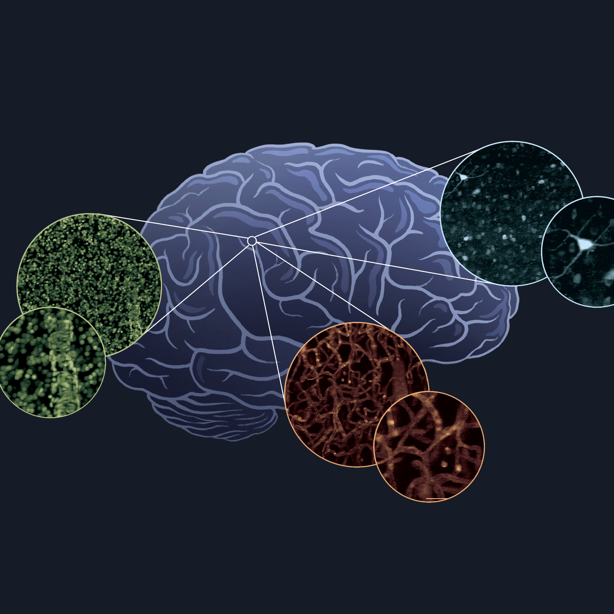
Bringing her engineering and physics expertise to neuroscience, Elizabeth Hillman has developed a wide range of multi-scale in-vivo imaging methods including SCAPE microscopy for high-speed 3D imaging of neural activity. She also uses these methods to understand blood flow in the brain as a way to improve human brain imaging.
Read more about Elizabeth Hillman, PhD >
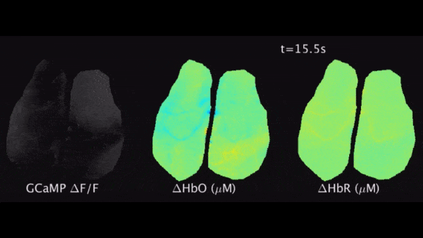
There is a crucial biological system that reaches every crevice of the brain, and yet goes unstudied by most neuroscientists. It is the vascular system, the network of vessels that carries oxygen-rich blood to hardworking nerve cells, called neurons. Elizabeth Hillman, PhD, a principal investigator at Columbia’s Mortimer B. Zuckerman Mind Brain Behavior Institute, studies neurovascular coupling — or how neural activity in the brain drives changes in brain blood flow. Despite its importance, little is known about this critical aspect of brain function.
“Studying the brain’s food supply has obvious connections to maintaining brain health,” Dr. Hillman says, “but understanding blood flow also holds the key to improving human brain imaging.” This is because one of our best tools for studying the human brain, a technique called functional magnetic resonance imaging (fMRI), measures blood flow as a way to detect patterns of brain activation. Yet debate over the relationship between neural activity and blood flow changes makes interpreting fMRI data very challenging.
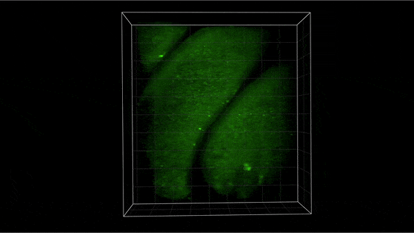
To study neurovascular coupling, physicist-turned-engineer Dr. Hillman has had to develop a range of unique imaging methods that go beyond fMRI to capture both neural activity and blood flow dynamics simultaneously in the living brain. In fact, developing new types of microscopes is a second important focus of her lab’s research program. Applying these techniques in a recent study looking at how neurons communicate with blood vessels, Dr. Hillman uncovered a sophisticated mechanism at play. She found that vascular endothelial cells, cells that line the insides of vessels, send electrical signals that tell the vessel to dilate and allow more blood through when neurons need it.
In addition to providing a clearer picture of the driving forces behind fMRI data, these results offer a new explanation for cognitive decline seen in conditions such as hypertension and diabetes, where endothelial cells are known to be impaired. Damaged endothelial cells, Dr. Hillman argues, could cause neurons to degrade — simply by restricting delivery of nutrients to hungry neurons. “Understanding these connections could yield fresh strategies to treat brain disorders,” she says, “while also revealing new ways to use techniques such as fMRI to diagnose and monitor disease in humans.”
Meanwhile, Dr. Hillman’s lab is home to a menagerie of advanced microscopes that she has developed to capture images of cells in action. One of her most exciting new inventions is SCAPE microscopy, which allows researchers to record three-dimensional, detailed videos of the brain at lightning-fast speeds: more than 100 times faster than competing technologies. “Every week we are finding that we can image new things, beyond anything we imagined,” Dr. Hillman remarks. This has included using SCAPE to capture neural activity throughout the brains of fruit flies and zebra fish, as well as image the brains of awake, behaving mice. The recipient of a recent award from the White House BRAIN Initiative, Dr. Hillman is currently commercializing SCAPE for widespread use. She hopes this technique will prove a game changer for neuroscience, but also that it have even broader impact as a tool for scientific and medical research.
Finding new ways to capture and interpret images is a passion for Dr. Hillman. “Imaging isn’t just about making pictures, it is about visualizing biology as it happens,” she says. “We are finally getting close to being able to answer so many questions about the brain.”
Project Lead (2017-present)
Adloph Lomb Medal (2011)
Fellow (2015)
Fellow (2017)
Fellow (2017)
Early Career Award (2006)
Young Investigator Award (2007)
Rodriguez Junior Faculty Award (2008)
CAREER Award (2010)
Voleti V, Patel KB, Li W, Perez Campos C, Bharadwaj S, Yu H, Ford C, Casper MJ, Yan RW, Liang W, Wen C, Kimura KD, Targoff KL,
Nat Methods.2019 Oct
Kozberg MG, Ma Y, Shaik MA, Kim SH,
J Neurosci.2016 Jun 22
Ma Y, Shaik MA, Kozberg MG, Kim SH, Portes JP, Timerman D,
Proc Natl Acad Sci U S A.2016 Dec 27
Galwaduge PT, Kim SH, Grosberg LE,
Biomed Opt Express.2015 Aug 1
Bouchard MB, Voleti V, Mendes CS, Lacefield C, Grueber WB, Mann RS, Bruno RM,
Nat Photonics.2015 Feb