NEW YORK, NY — Like a puppet pulled by strings, your body is controlled by brain cells called motor neurons. But how do each of these neurons get connected to the correct muscles in the first place?
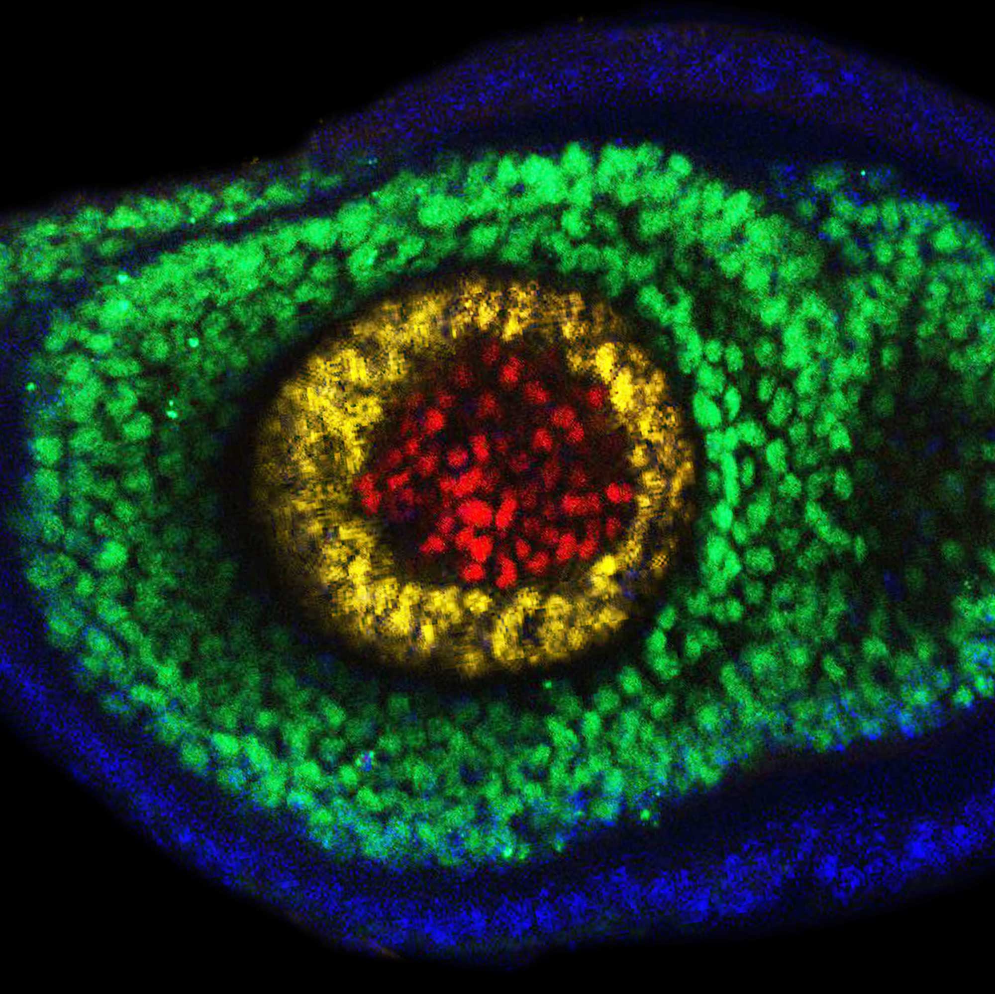
This video shows motor neurons (green) and muscle cells (pink) in the leg of a developing fruit fly embryo. Scientists at Columbia’s Zuckerman Institute found a pair of molecules that act like two halves of Velcro, helping the branches of such growing motor neurons properly stick to their corresponding muscle cells.
By mutating the proteins, called DIP-α and Dpr10, the researchers meticulously varied the strength of those neuron-muscle connections. ”Our goal was to understand the extent to which these two pieces of Velcro had evolved to interact with each other,” said doctoral student Davys Lopez of the Mann lab, first author on a paper detailing the discovery in Genes and Development.
When the interactions between these proteins are either too weak or too strong, the neurons fail to anchor correctly on muscles. This ultimately led the adult insects to have trouble walking.
"We are now developing new cutting edge techniques to help shed further light on these molecular interactions," said Richard Mann, PhD, the new study’s corresponding author, a principal investigator at Columbia’s Zuckerman Institute and the Higgins Professor of Biochemistry and Molecular Biophysics (in Systems Biology) at Columbia University's Vagelos College of Physicians and Surgeons. "This is just the beginning."