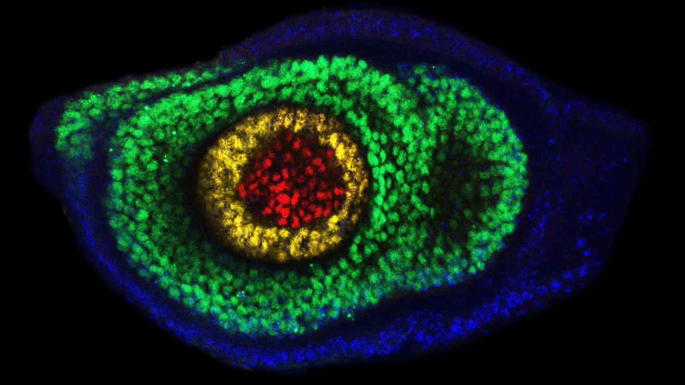
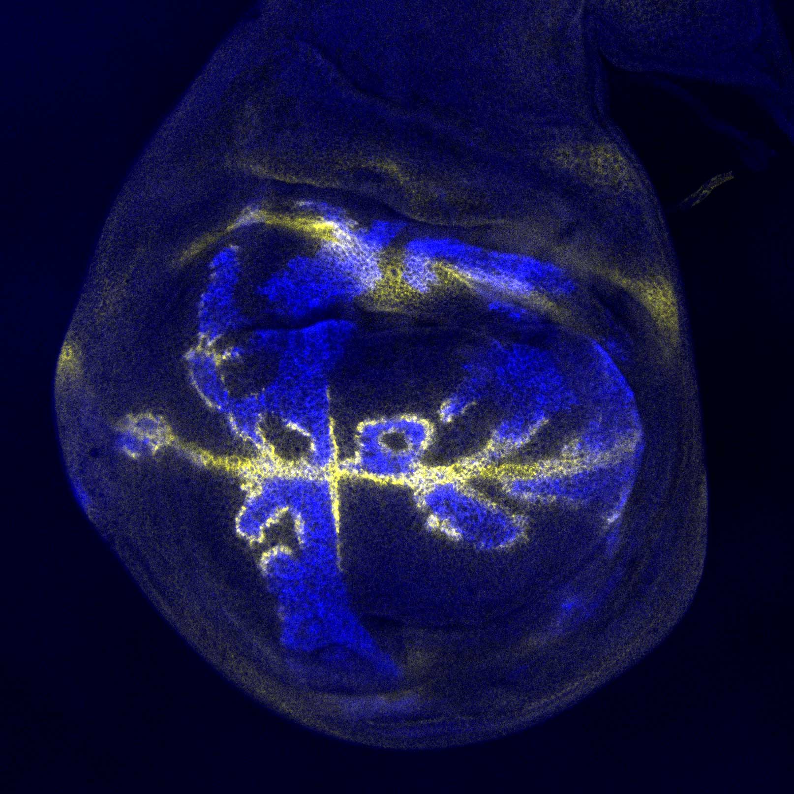
What do cancer and the growing legs of a fruit fly have in common? They can both be influenced by a single molecule, a protein that tends to call the shots inside of embryos as they develop into living, breathing animals. Present in virtually every creature on the planet, this protein goes by the name Epidermal Growth Factor Receptor protein, or EGFR.
Now a team of neuroscientists at Columbia University has figured out how to tease apart the many roles EGFR plays in the body — challenging conventional wisdom in the process. They report their findings in PLOS Genetics.
“The results of our research first and foremost solve a long-standing question about the nature of EGFR signaling and how it drives development,” said Richard Mann, PhD, a principal investigator at Columbia’s Mortimer B. Zuckerman Mind Brain Behavior Institute and the paper’s senior author. “But even more importantly, researchers can now use knowledge gleaned from our work to shed light on the link between EGFR and disease at a level of detail that until now would not have been possible.”
If we can use our enhancer method to systematically decode the regulation of EGFR activation, cancer researchers can then use that knowledge to identify potential targets for further development.
EGFR signaling is woven into the fabric of development. It guides the formation of many body parts and has been an intense area of research focus for decades. Indeed, EGFR is so critical to development that disruptions to its normal activity likely play a role in everything from developmental disorders to Alzheimer’s disease to cancer.
Scientists long believed that the molecules that bind to EGFR and activate it, known as EGFR ligands, act as morphogens. Morphogens are a type of signaling proteins that spread and trigger different responses in the developing tissues depending on their concentration — high levels result in one outcome while low morphogen levels lead to another response in the underlying cells.
“Morphogens coordinate the development of entire tissues from a single point source,” said Dr. Mann, who is also the Higgins Professor of Biochemistry and Molecular Biophysics at Columbia University Irving Medical Center. “Cells closest to a morphogen’s source get the largest, most intense concentration of signals, while cells farther away would get progressively less, like ripples in a pool.”
But some scientists have argued that EGFR signaling worked differently. Rather than sending out a single signal blast from a specific location, they said, multiple sources of EGFR ligands at different locations might send their own signals at different points of time — and this combination of signals together could guide development.
Distinguishing between these competing hypotheses is critical, the researchers point out. Without knowing how EGFR activation guides development, it is difficult to decipher what happens when the normal developmental process gets disrupted.
In addition, actually finding a way to test these two hypotheses has proven difficult. Scientists could not simply switch off EGFR signaling entirely and see what happens, as they often do to figure out how a protein affects development. Because EGFR signaling is so ubiquitous, too many other systems would be affected, to pinpoint exactly how it works.
To get around this problem, the Columbia team tried a new approach. They focused on the EGFR ligands’ many enhancers: small stretches of DNA that govern the ligands’ activity and trigger EGFR activity in a precise manner in different parts of the body.
“If you think of EGFR signaling as a soundboard, like what you’d find in a recording studio, enhancers are similar to the soundboard’s knobs and dials,” said Dr. Mann. “Just like turning those dials up or down changes the output of soundboard, turning up or down individual enhancers changes the ligand expression and, therefore, the resulting activity of signals.”
In this paper, the researchers first located the specific enhancers of the EGFR ligands that guided the fly’s leg development. They then switched off only those enhancers.
“In this way, we’ve managed to eliminate one small aspect of EGFR activity, leaving the rest of the signaling largely intact,” said Dr. Mann.
When doing so, they saw that the growth of the fly leg was not guided by a single source, the way a morphogen would act. Instead, the researchers found that EGFR sent signals from multiple different sources, located at different parts of the developing leg — knowledge made possible by the scientists’ focus on enhancers.
Because EGFR exists throughout the animal kingdom, these findings in the fly can be applied to studies of EGFR disruptions related to disease, such as developmental disorders and cancer.
“The overactivation of EGFR is associated with many different types of cancers, from small cell carcinoma to some types of breast and brain cancer,” said Dr. Mann. “If we can use our enhancer method to systematically decode the regulation of EGFR activation, cancer researchers can then use that knowledge to identify potential targets for further development of prognostic tests and therapies.”
###
This paper is titled: “Cis-regulatory architecture of a short-range EGFR organizing center in the Drosophila melanogaster leg.” Additional contributors include co-first and co-corresponding author Roumen Voutev, PhD, and co-first author Susan Newcomb, Aurelie Jory, PhD, Rebecca Delker, PhD, and Matthew Slattery, PhD.
This research was supported by the National Institutes of Health (R35GM118336 and RO1GM058575).
The authors report no financial or other conflicts of interest.


