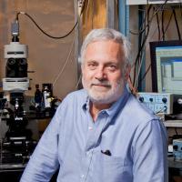
Gerald D. Fischbach, MD, Professor of Neuroscience and Professor of Pharmacology; Chair, Department of Neuroscience; Principal Investigator at Columbia's Zuckerman Institute
I wanted to uncover how something so tangible, like an electrical pulse, could represent something as complex and intangible as a memory.

When neuroscientist Steven Siegelbaum, PhD, first came to Columbia in 1981, his interests lay not in the mind — but in the heart.
“I spent my early career studying what regulates a heartbeat, work that I had intended to continue at Columbia,” said Dr. Siegelbaum, a principal investigator at the University’s Mortimer B. Zuckerman Mind Brain Behavior Institute. “But when I arrived, there was a mix up and my lab wasn’t ready.”
So Eric Kandel, MD, Nobel laureate and Zuckerman Institute codirector, offered up some space from his own lab.
“As I began working alongside Eric Kandel and members of his lab, I became fascinated by the intricacies of electrical activity in the brain,” said Dr. Siegelbaum. “I wanted to uncover how something so tangible, like an electrical pulse, could represent something as complex and intangible as a memory.”
This early work catalyzed a career that would take Dr. Siegelbaum on a journey deep into the brain — and reveal critical insights into how the brain forms and stores memories.
His first projects focused on the role of neural plasticity in memory. One of the key insights of modern neuroscience is that neural excitability and the process of neuronal communication is not fixed but can be altered as a result of experience. However, less was known about the underlying mechanisms of such changes. By examining neurons in the brain of the Aplysia, a marine snail, Dr. Kandel and colleagues found that the release of the brain chemical serotonin triggered long-term changes in neurons’ activity. And these changes, could be directly correlated to changes in the snails’ behavior. Dr. Siegelbaum believed the key was in the way neurons generated their electrical signals and how this information was passed on from neuron to neuron.
Neuroscientists had known that these electrical signals were mediated by the opening and closing of ion channels, specialized molecules in the neurons’ cell membrane that act as electrical gatekeepers, determining how and when a neuron generates an electric impulse.
However, the specific ion channel regulated by serotonin was unknown. Dr. Siegelbaum used a newly developed method, called the patch clamp technique, to identify that ion channel in Aplysia. He discovered that this ion channel, when exposed to serotonin, responded by shutting down for a prolonged period of time. Because this ion channel normally works by inhibiting neural activity, switching it off caused the neuron’s activity to ramp up. From the perspective of an Aplysia, this increased excitability of a neuron led to an enhanced response to a perceived threat. This simple mechanism represented the first record of a molecular event that was implicated in the encoding a memory.
“At the time, it was the closest thing to being able to watch the animal learn — in real time — at the level of individual molecules in neurons,” recalls Dr. Siegelbaum who is also professor and chair of the department of neuroscience at Columbia University Irving Medical Center.
Dr. Siegelbaum then turned his attention to the mammalian hippocampus, the brain region responsible for our ability to learn and remember new people, places, things and events. One goal in memory research is to explore whether and how different subdivisions of the hippocampus regulate these different components of memory. Siegelbaum became particularly interested in one small, relatively unexplored area of the hippocampus called the CA2 region.
“Through a combination of work in mice and in people, scientists have uncovered much about the role that different regions of the hippocampus play in memory,” said Dr. Siegelbaum. “But there was one glaring exception: CA2.”
Members of Dr. Siegelbaum’s lab developed a method to selectively turn off cells in the CA2 region in the brains of mice and then monitor changes in the animals’ memory.
The researchers discovered that the mice could still navigate a maze and recall inanimate objects and their surroundings, but were unable to recognize and recall fellow mice they had previously encountered. With their CA2 region of the brain altered, the mice had profound social memory deficits. These findings were published in April 2014 in Nature.
These results led Dr. Siegelbaum to speculate whether alterations in CA2 may contribute to social problems experienced by people with psychiatric diseases. In fact, evidence dating back to 1998 suggested that people suffering from schizophrenia and bipolar disorder showed a selective change in their CA2 region.
This prompted a long-term collaboration between Dr. Siegelbaum and Joseph Gogos, MD, PhD, an expert in the biology of schizophrenia and a fellow Zuckerman Institute Principal Investigator. In a 2016 Neuron paper, Drs. Gogos and Siegelbaum revealed a deterioration in the CA2 region in brain tissue from patients with schizophrenia and in mouse models with the disorder.
Now, the researchers are investigating whether drugs that target CA2 may help improve social memory in these mice, which may eventually lead to new treatments for patients.
“Our research into CA2 revealed a distinct brain region with a distinct purpose, and perhaps a distinct role in disease,” he said. “However, given the overall complexity of the brain, I suspect that CA2 is going to be involved in far more than just social memory.”
For example, unlike other parts of the hippocampus, CA2 contains high levels of oxytocin and vasopressin, two chemicals associated with social interactions such as bonding and aggression. Along with his collaborators, Dr. Siegelbaum is now studying the mechanisms by which CA2 cells respond to the release of these hormones. His latest results suggest that CA2 activity not only is important for social memory, it also promotes social aggression. When CA2 is turned off, a mouse is much less likely to attack an intruder mouse.
“The finding that the same small brain region controls both social memory and aggression came as a surprise,” said Dr. Siegelbaum. “Are memory and aggression inexorably linked, or are they two separate functions of the same brain region? And do changes to CA2 also contribute to changes in aggression observed in psychiatric diseases such as schizophrenia? These are questions I’m eager to investigate.”
Fellow (2015)
Member (2012)
Member (2012)
Srinivas KV, Buss EW, Sun Q, Santoro B, Takahashi H, Nicholson DA,
J Neurosci.2017 Feb 17
Masurkar AV, Srinivas KV, Brann DH, Warren R, Lowes DC,
Cell Rep.2017 Jan 3
Piskorowski RA, Nasrallah K, Diamantopoulou A, Mukai J, Hassan SI, , Gogos JA, Chevaleyre V
Neuron.2016 Jan 6
Basu J, Zaremba JD, Cheung SK, Hitti FL, Zemelman BV, Losonczy A,
Science.2016 Jan 8
Basu J,
Cold Spring Harb Perspect Biol.2015 Nov 2