NEW YORK, NY — Every moment we remember is thought to be encoded by changes in the strength of connections between brain cells, links called synapses. That’s been the theory for about half a century. But scientists had never actually imaged these synaptic changes happen as memories form in the brain of a complex living animal—until now.
In a paper published online today in Nature, researchers at Columbia's Zuckerman Institute have, for the first time, witnessed the memory encoding process as it occurs in mice.
"For decades, researchers who have studied how synapses are modified in the brains of mammals, or in the context of aging or neurodegeneration, psychiatric disorders or other conditions, have done so without having an actual readout at these vital links," said Franck Polleux, PhD, a principal investigator at Columbia's Zuckerman Institute. “Now we have the best view yet of how synapses change while a memory is forming.”
Throughout the brain, the electrical signals that transmit information are passed on from one neuron to another at synapses. Scientists had long thought the ability of synapses to strengthen or weaken over time, known as synaptic plasticity, helped govern whether memories last or fade.

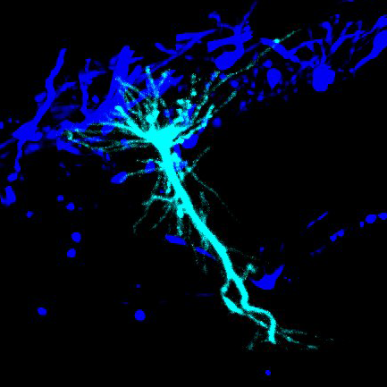
3D reconstruction of a single CA1 pyramidal neuron in the mouse brain. The dendritic arbor is in red and each yellow dot is a mapped excitatory synapse received by this neuron. Each CA1 pyramidal neuron in the mouse receives between 10,000 to 15,000 excitatory synapses. (Credit: Daniel Lascone / Polleux lab).
"Synaptic plasticity is the foundation for learning and memory," said Attila Losonczy, MD, PhD, a Zuckerman Institute principal investigator. "It's the basis of the human condition: how we learn, remember, adapt, and change our behavior."
Previous evidence for how synaptic plasticity might work emerged from experiments with relatively simple animals such as sea slugs, or under artificial conditions such as brain cells grown in lab dishes. Much remained uncertain about the actual way in which synapses change during learning in more complex animals such as mice and humans.
"The technical challenges of studying synaptic plasticity in a complex, living animal as they are awake and free to move, are immense," said Kevin Gonzalez, a graduate student in the Losonczy and Polleux labs and the study's first author. “That helps explain why nobody had been able to do this until now.”
In their new study, the researchers have overcome many of these technical challenges. They focused on the hippocampus, a key brain area for learning and memory. Pyramid-shaped neurons in the hippocampus’s CA1 region can become ‘place cells,’ which encode memories of locations. As individuals navigate around their environments, each place cell selectively fires at only one specific place.
We can track when during navigation each synapse is active and how strong they are, and we can do this for hundreds of synapses per neuron
Each CA1 neuron receives between 10,000 to 15,000 synaptic inputs from other neurons, each providing information about the entire space an animal is exploring. To help solve the mystery of how these hippocampal neurons each can use this barrage of information to become a place cell, the researchers genetically modified single CA1 neurons in the mouse brain to produce fluorescent molecules: red when the synapses activated, and green to display the strength of electricity flowing through them.
"We can track when during navigation each synapse is active and how strong they are, and we can do this for hundreds of synapses per neuron,” said Dr. Losonczy, who is also a professor of neuroscience at Columbia's Vagelos College of Physicians and Surgeons.
To visualize this synaptic light show, the researchers employed fast two-photon microscopy, a precise imaging technology.
"We're monitoring all this activity in structures one-hundredth the width of a human hair," Gonzalez said. “Our imaging technique is fast enough to capture changes in synapse strength that take hundredths of a second to happen.”
To image synapses at the moment of memory formation, the researchers spurred the genetically altered cells in the rodents to start encoding a memory of their current location as they explored a virtual environment. "This is a powerful approach. We don't have to wait for an event to occur that might generate a memory, we can now induce it when we want, and implant a single memory-encoding neuron in the brain,” Dr. Losonczy said.
Synapses that were active one to two seconds before the memory formed, about 3 to 5 percent of a neuron's synapses, became stronger over time, the researchers found. Synapses that were active outside this time window weakened.
"The brain boosts the strength of the synapses relevant to identifying a location and dampens others that are irrelevant," said Dr. Polleux, who is also a professor of neuroscience at Columbia's Vagelos College of Physicians and Surgeons.
This pattern of synaptic plasticity strongly supports the predictions of a recent model proposed by other researchers. This model helps explain how the brain can form a memory from a single event, as opposed to other models that require hundreds of experiences to produce a memory.
"These are the first experimental measurements of the rules of synaptic plasticity governing learning," Dr. Polleux said.
One of the biggest surprises for the scientists was their discovery that not all synapses in hippocampal neurons they monitored behaved in the same way. Neurons have branches like those of a tree: although the activity and strength of synapses in the branches near the apex of the pyramid-shaped cells changed in the experiments, those near the base of these cells did not.
"We still don't know why that is, or why it might be important," Dr. Losonczy said. "We know that memory is organized at multiple scales, from synapses to single neurons to neural circuits, and now we see that it may even be organized at a subcellular level."
Armed with the new results, "we are in a unique position to test the molecular basis of synaptic plasticity during memory formation, and with regards to potential defects underlying distortions or loss of memories in psychiatric or neurodegenerative diseases," Dr. Polleux said.
###
The paper, "Synaptic basis of feature selectivity in hippocampal neurons," was published in Nature on TKTK, 2024.
The full list of authors includes Kevin C. Gonzalez, Adrian Negrean, Zhenrui Liao, Satoshi Terada, Guofeng Zhang, Sungmoo Lee, Katalin Ócsai, Balázs J. Rózsa, Michael Z. Lin, Franck Polleux and Attila Losonczy.
The authors report no conflicts of interest.