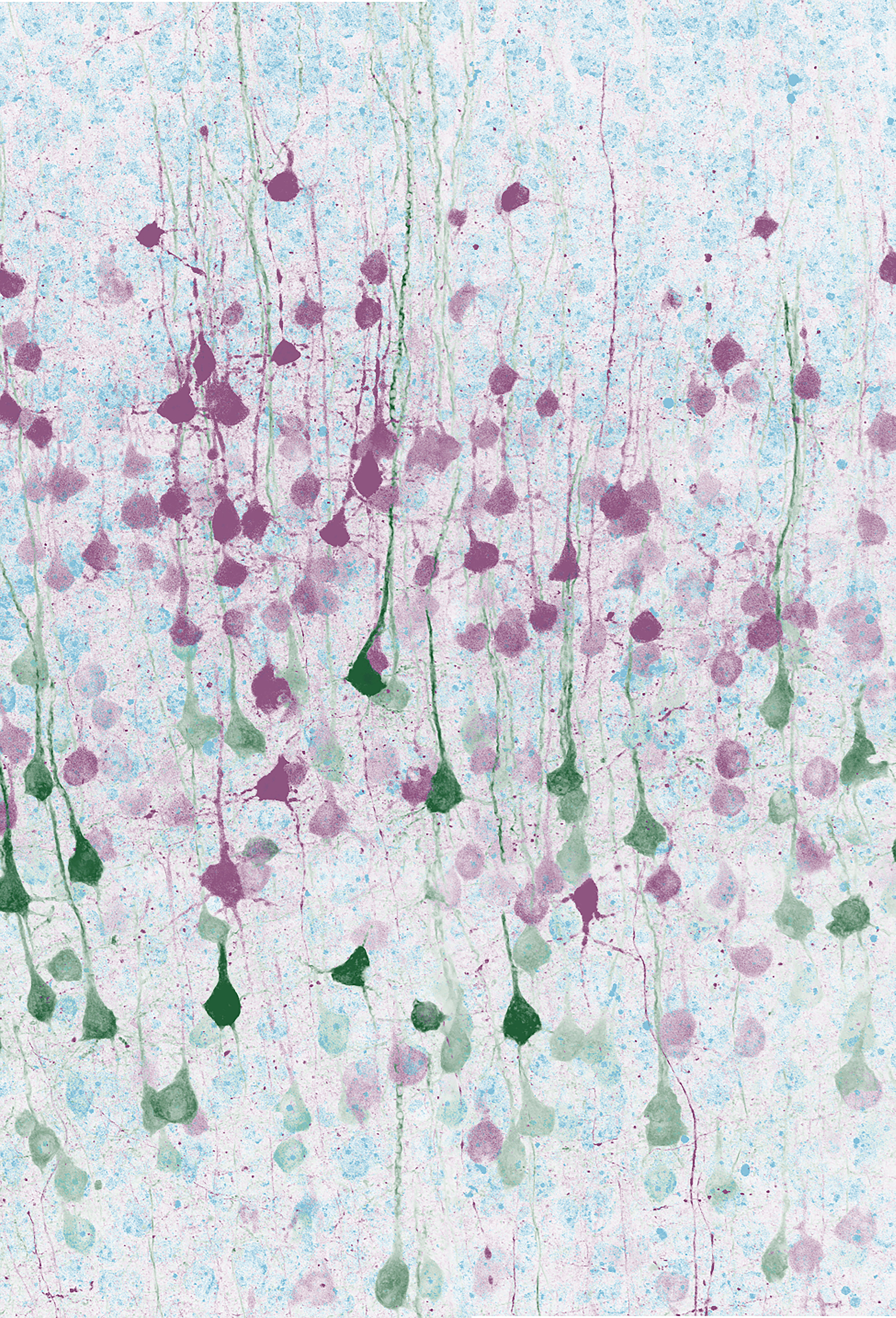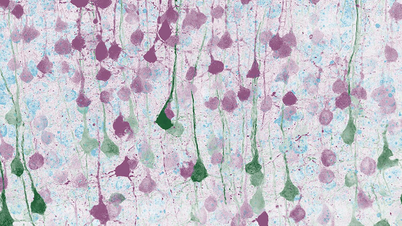
This may look like an abstract watercolor painting, but it’s actually an image from a microscope, showing nerve cells that help the brain to move the body. To create the image, postdoctoral research scientist Anders Nelson, PhD, injected a modified (and completely harmless) virus that can move from cell to cell and glows under fluorescent light. This technique reveals both the cells themselves and how the cells are connected to each other.
Dr. Nelson’s new experiments in the Costa lab explore how we learn to do complicated movements that require many steps. The nerve cells seen here carry different kinds of information from the brain to the body: including commands that make muscles twitch and the sequence of steps that must be followed to carry out an action. More specifically, the research image here shows two separate but intertwined neural pathways for shuttling this information: the corticostriatal neurons in purple, which connect two regions within the brain, and the corticospinal neurons in green, which connect the brain to the spine. Learn more about this research in the latest edition of Nature Neuroscience where it was recently published.